

700-924 | REM 4000 Endothelium Microscope
Precio habitual
Precio de venta
Precio habitual
Precio por unidad
Guarde
SKU: 700-924
700-924 | REM 4000 Endothelium Microscope
Precio habitual
Precio de venta
Precio habitual
Precio por unidad
Descripción
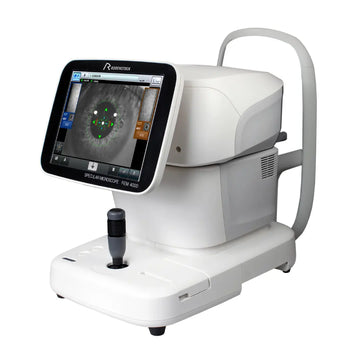
Rodenstock REM 4000 – Specular (Endothelium) Microscope
Stand-alone, fast and easy handling for reliable corneal endothelium analysis.
The REM 4000 combines non-contact imaging, auto-alignment, auto-measurement and integrated pachymetry in one compact unit. With 13 capture areas (centre + periphery) and automatic cell analysis (L-count, Core and Dark-area methods), you obtain reproducible endothelial metrics in seconds. A large 10.4” colour touchscreen, internal database and built-in thermal printer make the REM 4000 a true plug-and-touch solution for clinics and fitting rooms.
Key Benefits
- Fast & reproducible – Auto-alignment and auto-measurement deliver quick exams; automated capture of 16 images ensures consistent results.
- Comprehensive analysis – Automatic and manual modes with L-count, Core and Dark-area methods; counts up to 300 cells.
- 13 measurement areas – Centre plus 12 peripheral points increase diagnostic confidence, even with partial corneal opacity.
- Integrated non-contact pachymetry – Central corneal thickness measured automatically on every central exam.
- All-in-one workflow – 10.4″ touch UI, internal database (SD card), USB/LAN export and built-in thermal printer.
Specifications
| IMAGING & OPTICS | |
| Pixels used for picture taking | 480 (V) × 180 (H) pixels |
| Capturing scope (field on cornea) | 0.25 × 0.54 mm |
| Minimum cell resolution | 1.14 μm (V) × 1.45 μm (H) |
| Optical magnification | ×190 |
| Display | 10.4″ LCD colour touchscreen (display resolution 1.14 μm) |
| MEASUREMENT & FUNCTIONS | |
| Alignment / measurement | Auto-alignment, Auto-measurement; Manual mode 1 & 2 |
| Automatic capture | 16 images captured for analysis |
| Analysed cells (max.) | Up to 300 cells |
| Capture positions | 13 areas — centre + 12 peripheral points |
| Analysis methods | Automatic analysis, L-count, Core method, Dark-area method |
| Reported values | CD (cell density), AVG cell area, SD, CV, Cell size (max & min) |
| Integrated pachymetry | Non-contact; measured automatically on central exam; accuracy ±10 μm |
| Travel ranges | Moving section: X 88 mm, Y 40 mm, Z 50 mm; Electrical chin-rest stroke: 70 mm |
| DATA MANAGEMENT & CONNECTIVITY | |
| Built-in printer | Thermal printer (prints image and analysis) |
| Interfaces / export | USB-H ×2, USB-D ×2, LAN, SD card (internal database) |
| DIMENSIONS & ELECTRICAL REQUIREMENTS | |
| Dimensions (W × D × H) | 309 × 491 × 450 mm |
| Weight | Approx. 22 kg |
| Power supply | AC 100–240 V, 50/60 Hz |
| Power consumption | 100 VA |
| OPERATING ENVIRONMENT | |
| Temperature | +10° to +40° C |
| Humidity | 30% to 75% |
| Atmospheric pressure | 800 to 1060 hPa |
| Standards applied | MDD Annex II, ISO 13485 |